The term “congenital heart defect” refers to a disorder that occurs in utero, either in the anatomical structure of the heart, or in the vessels leaving/flowing into it, or in the valves located between the main cardiac cavities. There may be a combination of various defects with each other or with anomalies in the development of internal organs.
There are more than 100 types of congenital heart defects (CHD). Some of them increase the amount of blood going to the lungs, others reduce it, and others do not affect this indicator. In addition, each defect may have its own degree of severity, and this affects the course of the disease. Therefore, some anomalies of the heart and blood vessels are visible from birth and require emergency life-saving surgery, others are not so acute and are treated with medication (at least for a while). In some cases, heart defects appear suddenly and not in the first year of life.
Symptoms of heart defects range from minor to life-threatening. This is usually rapid breathing, the skin becoming bluish, poor weight gain, and fatigue when sucking. Chest pain is not typical for heart defects.
A little anatomy and physiology
This information will be useful to those who want to understand why a certain defect is more dangerous and why it has the symptoms it does.
The heart is an organ that performs the function of a pump. It is made up of four chambers - two atria and two ventricles. They are all made of three layers. The internal endocardium forms partitions between the heart chambers:
- Between the atrium and ventricle they look like valves. They open under pressure from blood entering the atrium to allow it to pass into the ventricle. After the blood flows into the ventricle, the valve flaps must close and prevent blood from flowing back into the atrium. Between the left chambers of the heart there is a bicuspid mitral valve, between the right chambers there is a valve consisting of three petals, which is called “tricuspid”.
- The two ventricles are separated by the interventricular septum - a rather dense structure, 7.5-11 mm thick, in the middle of which there is muscle tissue.
- The two atria are separated by the interatrial septum. It is thinner than the interventricular one, but, like the latter, it should not have any holes.
The aorta emerges from the left ventricle - the largest vessel with a diameter of 25-30 mm. The aorta, branching into many branches of smaller diameter, extending from it gradually, as internal organs appear along its path, carries oxygenated blood to them. Its last branches are the iliac arteries. They nourish the pelvic organs and also give off branches going to the legs.
“Wasted” blood, poor in oxygen, but with an abundance of carbon dioxide, leaves all internal organs through venules that flow into veins. The latter gradually also merge together:
- from the lower extremities, pelvis, abdomen they collect into the inferior vena cava (vena cava);
- from the arms, head, neck and lungs with the bronchi - into the superior vena cava.
Both vena cavae, above and below, flow into the right atrium. The blood makes a full circle in 23-37 seconds.
This is a large circle of blood circulation. In its arteries the pressure is higher than in the same vessels of the small circle.
The pulmonary circulation serves to ensure that all arterial blood in the systemic circulation can be oxygenated - saturated with oxygen.
It originates from the right ventricle, which pushes blood into the pulmonary trunk, which soon branches into the right (goes to the right lung) and left (goes to the left lung) pulmonary artery. Branching into smaller and smaller branches, the arterial vessels reach the alveoli of the lungs. There they give off carbon dioxide, which a person exhales.
Oxygen does not enter the arterial, but the venous blood of the pulmonary circulation. Venules with venous blood merge to form veins, and the latter, 4 in number, flow into the left atrium. The blood describes such a path (circle) in 4-5 seconds.
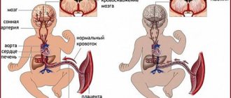
Placental circulation
While the fetus is developing in the uterus, its lungs are not used, since there is no connection between the baby and the surrounding air. But oxygen still reaches the baby, and this happens with the help of the placental circulation. It looks like this:
- oxygenated maternal blood enters intraplacentally;
- from the placenta it goes along the umbilical vein, which is divided into 2 parts:
- one goes into the inferior vena cava and mixes with the blood of the lower half of the body, which is already left without oxygen;
- the second goes to the portal vein, nourishes an important organ - the liver, and then mixes with the blood of the inferior vena cava;
- thus, venous-arterial blood flows through the inferior vena cava;
- non-oxygenated blood flows through the superior vena cava;
- From two vena cava, blood, like in a person after birth, enters the right atrium. But, unlike the extrauterine circulation, the right and left atria communicate with each other through the oval window;
- in the fetus, the foramen ovale is wide: almost all the blood from the right atrium enters the left, and then into the left ventricle;
- from the left ventricle blood enters the aorta;
- a small part of the blood goes from the right atrium to the right ventricle;
- from the right ventricle blood enters the pulmonary trunk;
- Since the lungs are collapsed, the pressure in the arteries that make up the pulmonary trunk is higher, so the blood has to be discharged into the aorta, where the pressure is still lower. This happens through a special vessel - the ductus botallus, which must close after birth. It flows into the aorta after the arteries to the head and upper extremities depart from it (that is, the latter receive more oxygenated blood);
- 60% of the systemic blood flow goes through the 2 umbilical arteries (they go on both sides of the umbilical vein) to the placenta;
- 40% of the blood from the systemic circle goes to the organs of the lower body.
It is precisely due to the structural features of the placental circle that the formed congenital heart defect does not lead to a significant deterioration in the child’s condition in utero.
Normal changes in the cardiovascular system after birth
When a baby is born, the foramen ovale, the communication between the atria, should close within the first year. This happens because when the lungs expand with air, the blood flow in the lungs increases. As a result, the pressure in the left atrium increases, and this “thrust” leads to the closure of the oval window. It does not heal immediately: the more the child screams, cries, and works on sucking (for example, with neurological problems or developmental defects such as a cleft lip), the longer a strong connective tissue – a “damper” – does not form in this place.
Regarding the ductus botallus, the situation is more complicated. It should close within the first 24 hours after birth, but if increased pressure remains in the pulmonary artery, it remains open. This is facilitated by hypoxia suffered during childbirth, which leads to spasm of the pulmonary vessels.
In the first 5-7 days, the pressure in the pulmonary artery decreases during heart contraction and should return to normal within 2 weeks after birth. Then it continues to decrease, because the pulmonary vessels undergo changes: the thickened muscle layer in them decreases, small pulmonary arteries disappear, some of the vessels straighten (previously they were tortuous). In addition, the alveoli, the main areas in which oxygen is exchanged between the air entering the lungs and the blood, expand in the lungs.
Incidence of congenital heart defects
Congenital heart disease in newborns occurs in 0.8-1.2%. In 2013, it was registered in 34.3 million people worldwide. It accounts for 10 to 30% of all congenital malformations and ranks second after malformations of the nervous system.
Depending on the nature of the diagnosis, congenital heart defects can be detected in 4–75 cases per 1000 infants born alive. In 0.6-1.9% they are moderate to severe. Congenital heart defects are the main cause of death in children from developmental defects. In 2013, for example, there were 323 thousand deaths worldwide, and in 1990 - 366 thousand. And if a child with a congenital defect lives to be 15 years old, then it is not necessary that he has “outgrown” it, and the risk of severe complications is now reduced.
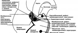
The most common heart defects are:
- ventricular septal defect (1/5 of all congenital defects);
- atrial septal defects (10-15% in the entire structure);
- open botal duct (10-15% in the entire structure);
- coarctation of the aorta;
- aortic stenosis;
- pulmonary stenosis;
- transposition of the great vessels.
There are defects that are more common in boys, others that are more typical for girls, but there are also those whose frequency is approximately the same in both sexes. Thus, in male infants, stenosis, coarctation of the aorta, transposition of the great vessels, common truncus arteriosus, tetralogy of Fallot, and pulmonary stenosis are more often found. In girls, patent ductus arteriosus, ventricular and atrial septal defects, and Fallot's triad are detected. Information about the gender of the developing fetus increases the likelihood of early diagnosis of characteristic defects.
The greatest concerns regarding the possible presence of such defects are in premature babies and those who were born weighing less than 3 kg. It is these newborns that require the prompt implementation of all diagnostic measures in order to identify possible developmental defects as early as possible.
Classification of defects
1. All congenital heart defects in children are divided according to the nature of the blood flow disturbance and the presence or absence of cyanosis of the skin (cyanosis).
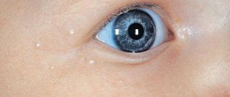
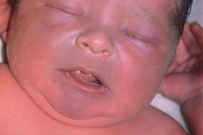
Cyanosis is a blue discoloration of the skin. It is caused by a lack of oxygen, which is delivered with the blood to organs and systems.
Personal experience! In my practice, there were two children with dextracardia (the heart is located on the right). Such children live normal healthy lives. The defect is detected only by listening to the heart.
2. Frequency of occurrence.
- Ventricular septal defect occurs in 20% of all heart defects.
- Atrial septal defect accounts for 5 - 10%.
- Patent ductus arteriosus accounts for 5 - 10%.
- Pulmonary stenosis, stenosis and coarctation of the aorta account for up to 7%.
- The remaining part is accounted for by other numerous, but rarer defects.
Why the heart can develop a birth defect
Often the cause of heart defects in newborns cannot be detected. In some cases, these may be individual reasons or a combination of them (most often a combination of genetic factors and various external influences):
Genetic factors
It can be:
- chromosomal disorders (in 5% of cases): trisomy on chromosomes 21, 13 and 18;
- gene mutations (in 2% of cases): in the genes TBX5, NKX2-5, TBX1, MYH6, GATA
Most often, these mutations are sporadic, occurring randomly and cannot be predicted before pregnancy. You can think about the fact that a child can be born with a congenital heart defect when there are (were) people in the family suffering from congenital heart defects, Down syndrome, Turner syndrome, Marfan syndrome, DiGeorge syndrome, Holt-Oram syndrome, Kartagener syndrome, Noonan syndrome and others. You also need to suspect a heart defect if a similar syndrome is detected in the fetus.
Infectious diseases
suffered by a pregnant woman, especially if it happened early in pregnancy. The most dangerous for the developing fetal heart are: rubella, viruses of the ARVI group (especially influenza and adenoviral infection), herpetic group (especially chicken pox and herpes simplex), viral hepatitis, cytomegaly, syphilis, tuberculosis, listeriosis, toxoplasmosis, mycoplasmosis.
Environmental factors
polluted air, radiation, living in mountainous areas or in places of high atmospheric pressure.
Toxic effects on the fetus if pregnant:
- takes certain medications: antibacterial and sulfa drugs, anticonvulsants and antiepileptic drugs (for example, Trimethadione), lithium preparations, painkillers and hormonal medications;
- smokes;
- takes drugs (amphetamines have a particularly toxic effect on the fetal heart);
- works with paint and varnish products, nitrates;
- drinks alcohol, especially in the initial weeks of gestation.
Causes related to maternal metabolism:
when she suffers from endocrine diseases (especially diabetes), is undernourished or, on the contrary, is obese.
If a pregnant woman is sick with diseases such as:
systemic lupus erythematosus, phenylketonuria, rheumatism.
At risk
In addition, we can say that the following pregnant women are at risk for developing congenital heart defects in children:
- over 35 years old or under 15 years old;
- having a history of stillbirth;
- with a history of spontaneous miscarriages;
- with bad habits (alcohol consumption is especially dangerous: the risk of congenital heart disease reaches 40%);
- if the child is conceived from a blood relative;
- when toxicosis of the first trimester is expressed;
- if the pregnancy proceeds with the threat of termination;
- whose family has relatives with heart defects.
Causes of congenital and acquired heart defects in newborns
Most congenital heart defects develop due to the adverse effects of the environment on the mother and fetus during pregnancy. Cardiologists include these types of influences:
- genetic risk factors;
- intrauterine infectious processes;
- age barrier of parents (over 35 for women and over 50 for men);
- environmental factors (radiation background, mutagenic agents, etc.);
- exposure to toxins (heavy metals, acids, alcohols, aldehydes, etc.);
- taking medications;
- mother's illness during gestation (severe toxicosis, diabetes mellitus, etc.).
The following children are at risk for congenital heart disease:
- with existing genetic diseases (including Down syndrome);
- born due to premature birth;
- with any other malformations of organs and systems.

Doctors call the main reasons for the appearance of acquired heart defects in newborns:
- streptococcal infection (due to rheumatic lesions of the body);
- severe hypertension;
- cardiomyopathy;
- toxic damage to the inner, middle and outer membranes of the heart;
- dysfunction of the conduction system of the main circulatory organ;
- injuries and tumors (in extremely rare cases).
At what age does heart disease develop?
The critical period when the above factors have a high chance of leading to the formation of heart defects is the first 3 months of pregnancy. At this time, cardiac structures are formed, and the influence of microbial, medicinal or industrial toxins on the fetal body can stop their development or lead to the formation of a heart “as at the previous stage of phylogenesis” (for example, as in reptiles, birds or amphibians). Cardiac structures are formed according to a clearly drawn up plan, and changes in it lead to the formation of one or another defect.
Around the 15th day of intrauterine development, the cells that give rise to the heart are located in the middle germinal layer (mesoderm) in the form of two horseshoe-shaped stripes. Some cells migrate here from a region of the outer germ layer (ectoderm) called the neural crest, a region that supplies different nerve cells to different parts of the body.
On day 19, 1 pair of vascular elements is formed - endocardial tubes. They merge with each other, the cells between them undergo programmed death, and the cells of the primary heart migrate to the tube and by day 21 form a ring of muscle cells around them. By day 22, the heart begins to contract and blood begins to circulate.
At the time of 22 days, the vascular system is a bilaterally symmetrical system with paired vessels on each side of the body and the heart, represented by a primitive tube located in the middle, in the middle layer of the body. Its sections, from which the atria are formed, are located further from the head (although it should be the other way around).
From days 23 to 28, the heart tube folds and twists. The future ventricles move to the left side of the center, taking their final location, and the atria move to the head end of the body. On the 28th day, the tissue of the heart tube expands, and within 2 weeks 4 heart cavities are formed here, separated by a primary membrane septum. If a damaging factor acts at this stage, blood will flow between the cavities of the heart.
Cells migrated from the neural crest give rise to the formation of the cardiac bulb, the main outflow tract from the heart. The bulb must be divided into 2 parts by the growing spiral septum, and then the ascending aorta and pulmonary trunk are formed. If the separation by the septum does not end, a defect is formed - a persistent ductus arteriosus. And if the vessels are located in the opposite direction, transposition of the great vessels results.
The two halves of the outflow tract must occupy certain positions at certain ventricles. When exposed to damaging factors at this stage, a “riding aorta” defect is formed (when the vessel originates from the interventricular septum).
Some of the cells of the septum primum die, forming an opening. At the same time, muscle cells grow here, forming a secondary septum, but the gap between the atria remains. This is the foramen ovale (window) - a shunt through which blood flows from the right to the left atrium. At the same stage, the ductus botallis is formed - a connecting channel between the aorta and the pulmonary artery.
What are the dangers of congenital heart defects?
Congenital cardiac defects lead to the development of major symptoms and complications through one of two mechanisms:
- The flow of blood through the vessels is disrupted. In case of anomalies with valve insufficiency or septal defects, the parts of the heart are overloaded with an increased volume of blood. If the defects involve narrowing (stenosis) of the openings or blood vessels, then the heart is overloaded with resistance. First, from any overload, the cardiac muscle layer increases, and the strength of its contractions increases. Afterwards, the compensation mechanisms “break down”, and the muscles of the enlarged heart become thinner. This disrupts systemic circulation and leads to cardiac failure.
- There is a disturbance in the systemic blood flow: either there is a lot of blood in the small circle, or there is little blood in one of the circles. Because of this, the supply of oxygen to organs deteriorates.
With “blue” defects, less oxygen is delivered due to a disruption in the speed of blood movement through the vessels. When the defect is “white,” hypoxia is associated with difficulties in the release of oxygen by hemoglobin molecules.
These pathogenetic mechanisms are the basis for the classification of congenital heart defects into phases, which, in turn, is used to determine treatment tactics. Thus, there are 3 phases of the disease:
Phase 1 – compensation and adaptation. The body compensates for the resulting disturbances by increasing the saturation of the myocardium.
Phase 2 is relatively compensatory. The heart muscle no longer functions as intensively. The structure and regulation of the heart is disrupted. The child’s physical development and motor activity improve.
Phase 3 – terminal. It occurs when the compensatory capabilities of the heart are exhausted, which is why dystrophic changes develop in the myocardium and internal organs. This stage ends with death. Infectious diseases, lung diseases and other pathologies can hasten its onset.
Types of Congenital Heart Defects
Congenital heart defects are most common in premature babies with low birth weight. The immediate causes of violations in the design of this organ are not fully understood. The coexistence of at least several genetic and environmental factors has a major influence on the occurrence of heart defects. The risk of their occurrence in a child increases when:
- A woman suffered from rubella in the first trimester of pregnancy.
- The pregnant woman smoked cigarettes, drank alcohol, or used drugs.
- Woman with diabetes.
- The child developed chromosomal abnormalities (including Down syndrome and Turner syndrome).
Heart defects can affect various structures of the organ, including the valves, ventricles, and blood vessels. Taking this into account it stands out:
- Defects in the parts of the heart.
- Abnormal abnormalities in large arteries.
- Improper blood flow to the heart.
- Partial or total stenosis of valves and blood vessels.
Classification of congenital cardiac defects
There are more than 100 forms of congenital heart abnormalities. Based on the nature of changes in blood circulation and, accordingly, the main symptoms, there are 2 main types of defects - “blue” (in which the baby’s skin has a bluish tint) and “white” (the baby’s skin is pale). They also have their own division.
"White" anomalies:
with them, arterial and venous blood do not mix. But this does not exclude the possibility of blood discharge from an area of higher pressure (from the left ventricle, that is, the systemic circle) to an area of lower pressure (to the right ventricle, the “source” of the pulmonary circulation):
- defects in which the volume of blood in the small circle increases. This is a functioning ductus arteriosus, atrial and interventricular septal defect, atrioventricular communication;
- anomalies associated with a decrease in the amount of blood in the pulmonary circulation: for example, isolated stenosis of the pulmonary trunk;
- defects that caused reduction (depletion) of blood in the systemic circle: isolated aortic stenosis, coarctation of the aorta;
- without any special discharge of blood from right to left. This occurs when the heart is structured normally, but is not located in its place, but on the right (dextrocardia), in the middle of the chest (mesocardia), in the abdominal cavity (abdominal dystopia of the heart), in the neck (cervical dystopia). A similar type of defect is characteristic of the bicuspid aortic valve (it should be tricuspid)
“Blue” defects, when mixing of arterial and venous blood occurs:
- when enrichment of the pulmonary circulation occurs (Eisenmenger syndrome, transposition of the great vessels);
- with depletion of the small circle: tetralogy of Fallot, Ebstein's defect.
Classification
Classification in medical practice of heart disease in a newborn is divided into 3 categories.
First category:
- Transposition (veins move to the place of arteries and vice versa).
- Tetralogy of Fallot (pathological enlargement of the right ventricle due to displacement of the aorta to the right side).
- Atresia (the lumen in the pulmonary artery in this case becomes overgrown).

Second category:
- Pathology of the septum, which is located between the atria.
- Pathology of the septum between the ventricles of the heart in a newborn.
Third category:
- Another type of congenital heart disease is stenosis (too narrow or too wide of the aortic valve).
- Insufficiency of one of the valves.
Any child can be born with defects; the main thing is to detect it in time and begin therapy. The classification of pathology varies according to symptoms.
How do congenital heart defects manifest?
Symptoms of heart disease depend on the type of pathology.
Signs of “blue” defects
Defects such as patent truncus arteriosus, tetralogy of Fallot, congenital fusion (stenosis) of the tricuspid valve, anomaly of the connection of the pulmonary veins are manifested by the following symptoms:
- the lips and nasolabial triangle may be bluish at rest (if the defect is significant), but this color can only appear during screaming, sucking or physical exertion;
- cyanotic fingers, which over time take on the appearance of “drum sticks”: completely thin, but thickened in the area of the nail phalanges;
- rough murmur over the heart;
- frequent infectious diseases, pneumonia;
- weakness;
- increased breathing;
- delayed physical and mental development;
- children are short;
- puberty occurs late.
In addition to changes in skin color, fatigue, weakness, shortness of breath, and changes in heart rate, congenital heart disease should be suspected if the child has one of these anomalies (abbreviation VACTERL):
- V – spinal anomalies (vertebral);
- A – anal atresia;
- C – cardiovascular (cardiovascular) abnormalities;
- T – transesophageal fistula (abnormal connections between the esophagus and other organs);
- E – esophageal (esophageal) atresia;
- R – renal (renal) anomaly;
- L – developmental defects of the limbs.
Signs of “white” defects
Such anomalies that occur with enrichment of the pulmonary circulation, for example, a ventricular septal defect, can be suspected immediately after the birth of a child. This:
- heart murmurs and increased heart rate, which are usually immediately heard by the neonatologist examining the baby immediately after birth;
- bluish tint of the skin, especially pronounced in the extremities;
- breathing that is more frequent than normal;
- poor appetite;
- light weight;
- lack of appetite or weak desire to breastfeed;
- inactivity;
- The baby sucks the breast/pacifier, but quickly gets tired and lets go.
If the septal defect is small, and all of these signs are mild, then poor weight gain should alert parents. Minor defects may even heal by the age of 10, but treatment should be carried out to reduce pressure in the pulmonary circulation system.
Anomalies of the interatrial septum are also characterized by disturbances in heart rhythm and the appearance of a protrusion of the chest wall in the area of the heart (“heart hump”).
An anomaly such as coarctation of the aorta is characterized by a feeling of heaviness and pulsation in the head, flushes of “heat” to the head, dizziness, and shortness of breath. Pulsating arteries are visible in the neck. Legs feel numbness, weakness in the legs; they are cold, and during physical activity there are cramps in the calf muscles.
If with a “white” defect there is no mixing of arterial and venous blood, this is manifested by the following signs:
- pain in the heart area;
- increased heart rate;
- dizziness, possibly with fainting;
- increased blood pressure;
- dyspnea;
- throbbing pain in the head.
Congenital heart defect
Approximately 1 in 100 children are born with a congenital heart defect. Congenital heart disease is the result of embryogenesis disorders.
Here are the causes of congenital heart defects:
- genetic factors: poor heredity, local gene changes and chromosomal mutations;
- Marfan syndrome;
- environmental factors, or mutagens: ionizing radiation, nitrates, phenols, excessive alcohol consumption, benzopyrene in smokers, antibiotics, non-steroidal anti-inflammatory drugs, etc.;
- maternal diseases: rubella, diabetes mellitus, systemic lupus erythematosus, cataracts, etc.
Congenital heart defects are divided into 2 groups: white and blue.
With white, or pale, heart defects, mixing of arterial and venous blood does not occur. White defects include: atrial septal defect, ventricular septal defect, AV communication, narrowing (stenosis) of the pulmonary artery, aortic stenosis, coarctation of the aorta, etc.
White heart disease in newborns, as a rule, does not manifest itself in any way. As they grow older, children with white heart defects develop clinical symptoms: pale skin, increased fatigue, shortness of breath, and retarded physical development.
Experiencing constant overload, the heart muscle wears out over the years - heart failure progresses: swelling of the legs appears, the liver enlarges, etc. Without surgery, the life expectancy of people with white defect is reduced by 20-25 years.
It should be emphasized that the presence of white congenital defects does not always provoke chronic heart failure. For a defect to manifest itself, it must be hemodynamically significant—the defect in the cardiac structure must be very significant. For example, an atrial septal defect of less than 1 cm (hemodynamically insignificant) in most cases does not manifest itself throughout life.
With blue heart defects, mixing of arterial and venous blood occurs. Examples of blue congenital heart defects: Eisenmenger complex, complete transposition of the great vessels, tetralogy of Fallot, Ebstein's anomaly, etc.
A typical symptom of blue heart disease is a bluish-gray, or cyanotic, color of the skin (cutaneous cyanosis) due to a lack of oxygen in the blood. All blue heart defects are hemodynamically significant; Cyanosis of the skin is clearly visible immediately after the birth of the baby.
Due to severe hypoxia of organs and tissues, blue congenital heart defects are much more dangerous than white defects - without surgical correction, the child may die in the first days and months after birth. Patients with blue defects without surgery live 30-50 years less than healthy people.
The most important condition for preserving the health and life of a child with congenital heart disease is timely diagnosis and surgical correction of the defect.
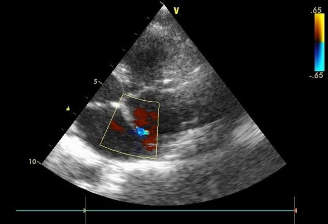
Congenital heart defects are diagnosed by a neonatologist (heart defects in newborns), family doctor, pediatrician (heart defects in children), therapist (congenital heart defects in adults), pediatric cardiologist, cardiologist, ultrasound specialist, endovascular surgeon, cardiac surgeon. The most accessible, safe and at the same time very informative instrumental diagnostic method is EchoCG with Dopplerography. To diagnose complex congenital heart defects, X-ray contrast studies (ventriculography, angiography), CT and MRI, including contrast, are used.
Congenital heart disease is diagnosed not only after birth, but also in utero using echocardiography with dopplerography. This is important for complex and incompatible defects. In the first case, pregnancy and childbirth are carried out taking into account the presence of a defect; they are planning cardiac surgery in the first days and months of life. In case of defects incompatible with life, artificial termination of pregnancy is offered.
In recent decades, almost all congenital heart defects have been successfully corrected surgically. A minimally invasive intervention (endovascular procedure) or a large open intervention, including the use of artificial circulation, is used. In the latter case, the heart is stopped and cut open, exposing the defect. In this case, the body is provided with blood and oxygen using a mechanical pump.
Postoperative mortality for most white and blue defects does not exceed 1%. With complex blue defects, in particular with complete transposition of the great vessels, postoperative mortality ranges from 5% to 50% depending on the level of the cardiac surgery center in which the surgery is performed.
Complications of congenital heart defects
The consequences of heart disease depend on the form of the defect and its severity. The main complications are:
- bacterial endocarditis;
- cardiac failure;
- frequent inflammation of the bronchi and lungs;
- attacks with shortness of breath and bluish skin;
- angina pectoris;
- myocardial infarction;
- thrombosis of peripheral veins;
- thromboembolism of cerebral vessels;
- rheumatic endocarditis;
- vascular aneurysms or their early atherosclerosis (typical of coarctation of the aorta).
Diagnostics
Some heart defects are detected during pregnancy using fetal echocardioscopy. This test is performed between 18 and 24 weeks of pregnancy using a transabdominal or transvaginal probe.
Often, congenital heart disease can be suspected after birth - by its characteristic appearance, rapid fatigue, and refusal to breastfeed. Sometimes the doctor’s attention is immediately drawn to disturbances in heart rhythm, heart murmurs, and expansion of the boundaries of this organ.
If no pathology is detected during examination, the defect can be suspected by a routine electrocardiogram or chest x-ray performed for suspected pneumonia. Heart defects are confirmed by echocardioscopy with Dopplerography (ultrasound of the heart with determination of blood flow in the cavities and through large vessels), but the final diagnosis is made in the cardiac center according to the following data:
- expert class echocardioscopy;
- inserting catheters into the cardiac cavities to measure pressure in them;
- angiocardiography.
Why does pathology develop?
Heart defects in newborns can be caused by various factors. Most of them cannot be influenced even with the help of medicine.
Why does congenital heart disease develop in newborns?
- Chromosome mutations at the genetic level as a cause of heart defects in newborns.
- Infectious diseases in women during pregnancy (rubella, toxoplasmosis, influenza and many others). They can cause particular harm if a woman falls ill in the first trimester, when all the organs of the embryo are formed.
- Chronic diseases such as diabetes, lupus and others.
- In rare cases, the cause is the age of the parents.

- Living in an unfavorable ecological environment.
- Using medications without a doctor's permission.
- Bad habits (alcohol, smoking, drugs).
- Psychological discomfort of a pregnant woman. If a woman has had miscarriages or a frozen pregnancy, then in subsequent pregnancies she may be constantly stressed from the experience.
- Heredity. Heredity plays a major role in the development of heart disease in children. If there are people in the family who suffer from a similar disease, then the child may be born with the pathology.
The causes and consequences of heart pathology in each individual baby can be varied.
Therapy of defects
Treatment of congenital heart defects is divided into medical and surgical. The first type is used for minor defects, as well as at the stages of preparation for and after intervention. It is aimed at stabilizing pressure in the pulmonary artery system or systemic circulation, improving myocardial trophism and oxygen absorption by internal organs. For this purpose they prescribe:
- diuretics;
- potassium salts;
- digitalis preparations;
- antiarrhythmic drugs;
- beta blockers;
- indomethacin - according to the regimen. This is the only radical drug treatment that can completely solve the problem in the case of an open ductus arteriosus.
Some cardiac anomalies can resolve on their own, without surgical treatment, but this usually does not apply to “blue” defects.
Surgery for heart defects takes into account the type and phase of the defect:
- If the cardiac defect is within phase I, emergency surgery is performed before one year of life. For example, when the small circle is depleted, this is artificial stenosis of the pulmonary artery; when the small circle is overfilled, it is the application of an artificial ductus arteriosus.
- In phase II, a planned operation is carried out, which is carried out after careful preparation, at various times (usually before puberty).
- In case of decompensation, when the defect is detected already in the third phase, only this type of intervention can be carried out that will slightly improve the child’s quality of life.
If the cardiac anomaly was discovered before birth, then some types of operations can already be performed in utero. In cases where this is not possible and the defect is a life-threatening anomaly, the woman is given birth in a specialized hospital, after which the child undergoes the necessary intervention immediately after birth. For some serious defects, a heart transplant may be possible.
Forecast
Congenital heart disease - how long do you live with it? This is based on the form of the anomaly:
- With a functioning ductus botallus, septal anomalies or stenosis of the pulmonary artery, mortality in the first year of life without treatment is 8-11%.
- Tetralogy (a combination of 4 defects) of Fallot and pathologies of the myocardium structure cause a mortality rate of 24-36% in children under one year of age.
- Coarctation, aortic stenosis and aortic dextraposition lead to 36-52% mortality in the first year of life. Average life expectancy is 12 years.
- With left ventricular hypoplasia, pulmonary atresia, and common aortic trunk, 73-97% die in the first 12 months of life.
To prevent the formation of cardiac defects as much as possible, even before pregnancy, a woman should be vaccinated against rubella, add iodized salt and folic acid to her food. While pregnant, the mother should not smoke, take alcohol or take drugs. If there are frequent cases of heart defects in the family, a woman or man should consult a geneticist and, perhaps, not plan a pregnancy.
Author:
Krivega Maria Salavatovna resuscitator
Symptoms and treatment of congenital heart disease in newborns
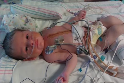
Congenital heart disease in newborns (CHD) is a serious anatomical defect that occurs even at the stage of intrauterine development of the fetus. There are many reasons why children can be born with this pathology. Many parents do not know what to do when making such a diagnosis. Experts divide the disease into several types according to degree of complexity. There are “white” and “blue” heart defects, as well as defects of the heart valves. In the first case, the problem lies in the valve partitions, in the second - in the vessels, and in the third - in the valves.

Heart defects in newborns are not uncommon. About a third of babies born with congenital heart disease die from this terrible disease. But in some cases, everything can be corrected with surgery. Without surgery, the child has little chance of survival. About 70% of babies with heart defects who do not have surgery on time die in the first year of life.
Finding out the reason why such a pathology appears is quite difficult. This can be influenced by various factors, including the mother’s poor lifestyle and illnesses suffered during pregnancy. There are several groups of people who may have a baby with congenital heart disease.
- bad heredity
- chromosome disorder
- gene mutation
- effect of harmful substances