How does the size of the uterus change?
During pregnancy, the uterus changes not only its size, but also its consistency, shape and location. At 5-6 weeks of pregnancy, the body of the uterus grows slightly. First, it takes on the shape of a ball due to an increase in the anterior-posterior size, then an increase in the transverse size is observed. In early pregnancy, asymmetry of the uterus is very often observed, in which one of its corners protrudes. It depends on where the fertilized egg is attached to the endometrium. As it grows, the uterine cavity is completely filled, and the asymmetry disappears. At the 8th week of pregnancy, a twofold increase in the uterus is observed, and at the 10th week – threefold. At the 12th week, the fundus of the uterus reaches the plane of exit from the pelvis, that is, it is located on the upper edge of the symphysis pubis.
Uterus: a little physiology
The uterus is a unique organ, the structure of which is such that it is capable of stretching and increasing its size tens of times during pregnancy and returning to its original state after childbirth.
The uterus has a larger part, the body, located above, and a smaller part, the cervix. Between the body and the cervix there is an intermediate section called the isthmus. The highest part of the uterine body is called the fundus. The wall of the uterus consists of three layers: the inner - endometrium, the middle - myometrium and the outer - perimeter (serous membrane).
The endometrium is a mucous membrane that changes depending on the phase of the menstrual cycle. And if pregnancy does not occur, the endometrium is separated and released from the uterus along with blood during menstruation. If pregnancy occurs, the endometrium thickens and provides the fertilized egg with nutrients in the early stages of pregnancy.
The main part of the uterine wall is the muscular layer - the myometrium. It is due to changes in this membrane that the size of the uterus increases during pregnancy. The myometrium consists of muscle fibers. During pregnancy, due to the division of muscle cells (myocytes), new muscle fibers are formed, but the main growth of the uterus occurs due to elongation by 10-12 times and thickening (hypertrophy) of muscle fibers by 4-5 times, which occurs mainly in the first half of pregnancy By the middle of pregnancy, the thickness of the uterine wall reaches 3-4 cm.
length - 37-38 cm, anteroposterior size - up to 24 cm, transverse size - 25-26 cm. The weight of the uterus by the end of pregnancy reaches 1000-1200 g without the child and membranes. With polyhydramnios or multiple pregnancy, the size of the uterus can reach even larger sizes. The volume of the uterine cavity increases 500 times by the ninth month of pregnancy.
How the height of the uterine fundus changes by week
At the 14th week of pregnancy, when the uterus extends beyond the pelvis, the obstetrician-gynecologist begins to regularly measure the height of the uterine fundus. The height of the uterine fundus (UFH) is considered to be the distance between the upper edge of the symphysis pubis and the outermost point of the uterus. At 16 weeks, AMD is 6-7 cm, and the location of the organ is the level between the navel and the symphysis pubis. At 20 weeks, the size of the AMD varies from 12 to 13 cm, and the uterus is located 2 cm below the navel. At 24 weeks, the organ reaches the level of the navel, and at 28 weeks it exceeds it by 2-3 cm. At 32 weeks, the uterus stops in the middle between the navel and the urinary process, that is, in the lower part of the sternum.
At 36 weeks, the peak growth of the uterus is observed, with the organ reaching the costal arches and the urinary process. At the end of pregnancy, the fundus of the uterus drops slightly. In general, the displacement of the uterus during pregnancy can be characterized as follows: for the first three months it is located in the abdominal cavity, in the 4th month it reaches the level between the navel and pubis. In the 5th month, the location of its bottom is the level of the navel, and by the end of pregnancy the bottom reaches the lower edge of the sternum. In the last weeks, the uterus rises so high that it puts pressure on the diaphragm and other abdominal organs - the bladder, stomach and intestines, creating for the woman many problems associated with constipation, heartburn, frequent urination, breathing difficulties, etc.
How the uterus enlarges by week of pregnancy
The uterus is an important female reproductive organ, which is responsible for bearing a child. It is in this environment that the implantation of the embryo and its development occur. The location of the reproductive organ is in the pelvic area.
A woman seeks help from a gynecologist when unusual abnormalities appear in the menstrual cycle or when planning pregnancy.
When diagnosing a problem, a specialist may resort to the following examinations:
- Manual examination;
- Examination using a gynecological speculum;
- Smear analysis for vaginal microflora;
- Analysis of material to identify viruses and infections;
- Colposcopy;
- Ultrasound examination;
- Hysteroscopy and laparoscopy;
- Taking blood for hormone analysis.
- Girls under 1 year of age have a uterus size of up to 4 cm.
- Afterwards the organ is reduced in size by 2 times.
- The uterus remains in this state for up to 7 years.
- Afterwards, until puberty, it increases and stabilizes.
- Subsequently, serious changes in the uterus occur with pregnancy. Then it stretches and contracts after childbirth. Small increases before pregnancy may occur for normal reasons.
- Menopause or menopause in women also causes changes in the size of the uterus. During this period, female sex hormones, progesterone and estrogen, are not able to be produced by the body in the required quantities.
When specialists, based on ultrasound results, observe small indicators of the size of the uterus, they indicate the presence of a small reproductive organ.
There are several stages when deviation from the norm may appear in a reduced form:
- The hypoplastic stage indicates a normal state of the uterus, which corresponds to adolescence.
- The infantile stage usually manifests itself in a child, but if it occurs in an adult woman, this indicates a deviation.
- The aplasia stage manifests itself in a teenager, the uterus develops like that of a small child.
When the reproductive organ is underdeveloped, the following symptoms may occur:
- Menstruation came at the age of 16 years;
- Late puberty;
- Girls suffer from signs that are not typical for their body;
- There is no menstrual cycle or it is not constant;
- Conception cannot be achieved for a long time.
A small uterus is a problem that is associated with underdevelopment of the reproductive organs. In order to conceive, a woman will have to take a lot of effort.
When experts discover deviations from the normal size of the uterus in women, this negatively affects their health.
An enlarged condition may be normal in the following cases:
- When does puberty occur?
- The moment of the beginning of pregnancy.
- After childbirth, for some time.
When a woman has an enlarged uterus, this may indicate the occurrence of pathological processes.
This condition can be caused by:
- Uterine fibroids cause benign formations.
- A uterine cyst is a pathological formation in its tissues.
- Formations in the form of cancerous tumors.
- Proliferation of the mucous membrane of the uterine cavity.
- Disturbances in the endocrine system.
During this time, women experience the following symptoms:
- Feeling of pain in the lower abdomen.
- Frequent urination or urinary incontinence.
- Disruption of the menstrual cycle, and they become abundant.
- The total weight increases.
- Headaches occur.
During an ultrasound examination and detection of pregnancy, the size of the reproductive organ will increase. The main indicator at this moment is the length of the uterus.
The gynecologist monitors her indicators starting from the 3rd trimester. Before this period, the size of the uterus is checked by manual examination or ultrasound examination using a vaginal probe.
A specialist is able to determine the gestational age by the size of the reproductive organ. He is able to detect abnormalities using the size of the uterus. The gynecologist is able to use palpation to determine how the organ will increase further.
This happens when a woman begins her 3rd month of pregnancy. With each week, 1 cm is added to the previous size. At the time of birth, this figure can reach up to 40 cm.
If a slight deviation from the norm occurs, this is not a pathology in all cases. The indicators for each woman are individual and if they are good enough, the specialist recommends undergoing a preventive examination once a year.
However, for a number of reasons, the growth of the uterus may slow down, which leads to the occurrence of a pathology - an infantile (children's) uterus. Its length is about 3 cm. Along with the small size of the uterus, the ratio of the lengths of the body of the uterus to the cervix changes, which, in contrast to the normal one (2/1), is equal to 1/3.
A child's uterus (infantile uterus or uterine hypoplasia) is an anomaly characterized by underdevelopment of the female reproductive system. In this case, the uterus is correctly formed, but after the birth of a girl it lags behind in development.
The formation of a “baby” uterus may be associated with a disorder of embryonic development or caused by external factors affecting the girl after her birth, such as:
- imbalance in the regulation of the reproductive system and decreased estrogen synthesis;
- stress, mental disorders, depression;
- sports overload;
- cruel diets leading to the development of anorexia;
- infectious and viral diseases (ARVI, influenza, tonsillitis and others);
- intoxication: smoking and drugs.
The normal size of the uterus in women differs depending on age, number of pregnancies, abortions and hormonal levels. Specialists measure the length and width of the uterus and indicate the thickness of the walls.
Women of childbearing age have a uterus of the following sizes:
- length – up to 70 mm;
- width – up to 60 mm;
- wall thickness – up to 42 mm.
If a woman has not given birth or become pregnant, the length of the uterus is 47 mm; in those who have given birth, the figures increase to 61 mm. Moreover, with each pregnancy the length of the uterus and its width increase. When there were pregnancies, but without childbirth, the length of the organ will be within 54 mm.
To obtain accurate results, it is recommended to do an ultrasound on days 5-8 of the cycle, and repeat examinations at the same phase. The conclusion may be affected by hormonal fluctuations, the presence of pregnancies and childbirth. Sometimes even stress and nervous overload lead to changes in size.
We suggest you read: The mucus plug came away after examination by a gynecologist
During menopause, the production of female sex hormones decreases, which causes a decrease in the size of the uterus. At the beginning of postmenopause, the length of the uterus varies from 40 to 70 mm, width - 27-54 mm, and wall thickness - 18-36 mm. 15-30 years after the start of changes, the length of the uterus will be 33-67 mm, width - 25-54, wall thickness - 14-36.
Sizes can vary greatly, as it all depends on the time of onset of menopause and the individual characteristics of the body. For some women, menopause begins before the age of 40, and for others it occurs after 60. If the indicators do not differ much from the norm at the time of the onset of menopause, there is a high probability of becoming pregnant through artificial insemination.
The overall size of the uterus affects the length of its cervix. In women of childbearing age, the length of the cervix does not exceed 4.5 cm, and the thickness is 0.3 cm. If the body and cervix are bent, the thickness can reach 4.5 cm, which is a pathology.
Numerous changes occur in the body of the expectant mother that allow her to bear a child. But there is no doubt that the most significant changes occur in the uterus - the organ in which new life develops.
Pregnancy is characterized by an increase in the size of the uterus, a change in its consistency (density), and shape.
Enlargement of the uterus begins at 5-6 weeks of pregnancy (with 1-2 weeks of delay), while the body of the uterus increases slightly. First, the uterus increases in anteroposterior size and becomes spherical, and then the transverse size also increases. The longer the pregnancy, the more noticeable the enlargement of the uterus becomes.
In the early stages of pregnancy, asymmetry of the uterus often occurs; during bimanual examination, a protrusion of one of the corners of the uterus is palpated. Protrusion occurs due to the growth of the fertilized egg; as pregnancy progresses, the fertilized egg fills the entire uterine cavity and the asymmetry of the uterus disappears. By 8 weeks of pregnancy, the body of the uterus increases approximately 2 times, by 10 weeks - 3 times. By 12 weeks, the uterus enlarges 4 times and the fundus of the uterus reaches the plane of exit from the small pelvis, i.e., the upper edge of the symphysis pubis.
From the second trimester of pregnancy (from the 13-14th week of pregnancy), the uterus extends beyond the pelvis and can be palpated through the anterior abdominal wall. Therefore, starting from this period, the obstetrician-gynecologist measures the height of the uterine fundus (VDM - the distance between the upper edge of the symphysis pubis and the highest point of the uterus) and abdominal circumference.
All measurements are recorded in the pregnant woman’s individual chart, which allows you to track the dynamics of uterine growth and assess the growth rate. VMR is measured with a centimeter tape or a pelvisometer (a special device for measuring the distance between two points) with the pregnant woman lying on her back. Before measuring and examining, you must empty your bladder.
The normal (physiological) course of pregnancy is characterized by the following indicators of AMR:
- at 16 weeks of pregnancy, the fundus of the uterus is located in the middle of the distance between the navel and the pubic symphysis, the IMD is 6-7 cm;
- at 20 weeks, the fundus of the uterus is 2 cm below the navel, IMD 12-13 cm;
- at 24 weeks, the fundus of the uterus is at the level of the navel, UMR 20-24 cm;
- at 28 weeks, the fundus of the uterus is 2-3 cm above the navel, VDM - 24-28 cm;
- at 32 weeks, the fundus of the uterus is in the middle between the navel and the xiphoid process (the lower part of the sternum), the IMD is 28-30 cm;
- at 36 weeks, the fundus of the uterus rises to the xiphoid process and costal arches. During this period, the highest position of the uterine fundus is observed. VDM - 32-34 cm;
- at the end of pregnancy (at 38-40 weeks), the fundus of the uterus descends, the IMD is 28-32 cm.
At the same time, the height of the uterus is affected by the size and position of the fetus, the amount of amniotic fluid, and multiple pregnancies. With a large fetus, twins, or polyhydramnios, the uterus stretches more, and accordingly, the fundus of the uterus will be higher. With oblique or transverse IMD may be less than normal. It should also be noted that the height of the uterine fundus at the same stage of pregnancy in different women fluctuates by 2-4 cm due to individual characteristics, therefore, when determining the duration of pregnancy, they are never guided only by the size of the uterus. Other indicators are also taken into account, such as the date of the last menstruation, the date of the first fetal movement, and the results of an ultrasound examination.
- Until 12 weeks of pregnancy, no changes occur in the abdomen. Throughout the first trimester, the tiny embryo is reliably protected from external influences by the bones of the small pelvis.
- From the 12th to the 16th week of pregnancy, an enlarged abdomen can only be determined by the experienced eye of a friend, and only if the pregnant woman is thin.
- From the 17th to the 20th week of pregnancy, the belly becomes noticeable to others if the woman wears tight-fitting clothes. Usually it is at this time that the expectant mother first thinks about updating her wardrobe.
- After the 20th week, pregnancy is already difficult to hide from others. This can only be done by overweight women or those who wear overly loose clothing.
- After the 28th week of pregnancy, the second period of wardrobe renewal begins, as the growing belly requires special clothing. Only a few overweight women manage to hide their pregnancy.
How many weeks does the uterus rise during pregnancy?
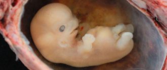
However, already at 10 weeks the uterus becomes crowded in the pelvic area. By this time, the baby already has arms, legs, tiny fingers and even nails on them. His ears and upper lip are clearly visible, a diaphragm appears, and the rudiments of baby teeth are formed. During this period, changes occur in the red blood cells of the fetus, agglutinogens responsible for determining the blood group begin to appear. Despite its miniature size, the future little man is already fully formed by this time, and all that will happen to him next is rapid growth. If by the beginning of the 11th week no abnormalities in the development of the fetus have occurred, we can say with confidence that the baby will not have any congenital pathologies.
Uterus during pregnancy
During pregnancy, the uterus grows and stretches continuously in size, parallel to the growth of the baby in it. A gradual displacement of the uterus also occurs: if during the first three months the uterus is located in the abdominal cavity during pregnancy, in the 4th month its bottom reaches the level between the navel and the pubis, by the 5th month the bottom is determined at the level of the navel, and in the later stages of pregnancy - lower edge of the sternum. Towards the end of pregnancy, the uterus rises so high that it puts pressure on the diaphragm, making it difficult for the mother to breathe. At the same time, constantly increasing in size, the uterus during pregnancy also puts pressure on the abdominal organs: it compresses the stomach and intestines, and the bladder. This explains the frequent cases of constipation, heartburn, digestive difficulties, and frequent urination during pregnancy.
How the belly grows during pregnancy
the growth of the fetus in the uterus and the increase in the amount of amniotic fluid at each appointment of a pregnant woman using external obstetric examination techniques. The woman lies on her back, legs straightened, and the bladder must be emptied before the examination. The doctor measures the distance from the upper edge of the symphysis pubis to the most prominent part of the uterine fundus with a centimeter tape, determining the height of the uterine fundus (UF), as well as the abdominal circumference at the level of the navel (UF). The measurements help you figure out the rate at which your belly is growing. Approximately, the height of the uterine fundus in centimeters corresponds to the gestational age in weeks. Abdominal circumference depends on many factors, primarily on the woman’s constitution and her weight. An increase in abdominal circumference and weight gain are directly related. On average, abdominal circumference changes by 1 cm each week starting at 20 weeks of pregnancy .
Video about normal uterine sizes
After the birth of a child and placenta, already in the first hours of the postpartum period, a significant contraction (reduction in size) of the uterus occurs. The height of the uterine fundus in the first hours after birth is 15-20 cm. Restoration of the uterus after childbirth is called involution. During the first two weeks after birth, the fundus of the uterus drops approximately 1 cm daily.
- On the 1-2 days after birth, the fundus of the uterus is at the level of the navel - IMD 12-15 cm;
- on the 4th day of VDM - 9-11 cm;
- on the 6th day of VDM - 9-10 cm;
- on the 8th day of VDM - 7-8 cm;
- on the 10th day of VDM - 5-6 cm;
- on the 12-14th day, the fundus of the uterus is located at the level of the junction of the pubic bones.
The uterus completely shrinks to the size it was before birth in approximately 6-8 weeks. Reverse development of the uterus depends on many different factors: the characteristics of pregnancy and childbirth, breastfeeding, the woman’s age, general condition, and the number of births in the anamnesis. The uterus contracts more slowly in women over 30 years of age, in weakened and multiparous women, after multiple pregnancies and pregnancies complicated by polyhydramnios, with myomema, as well as when inflammation occurs in the uterus (endometritis) during pregnancy, childbirth or the postpartum period.
Marina Ershova, obstetrician-gynecologist, Moscow
So, let's find out what affects the increase in abdominal parameters, how it grows week by week, how its growth differs during multiple pregnancy and why the stomach needs a bandage.
Another parameter that influences the onset of the appearance of a tummy is the woman’s activity. If she plays sports or is a professional athlete, then the roundness of her tummy will appear by the third trimester. And then when wearing tight, tight clothes. It is a mistaken belief that the roundness of the abdomen depends on the size and weight of the fetus. Little actually depends on these factors. After all, all babies in the mother’s womb grow and develop approximately the same until the third trimester.
It should be borne in mind that abdominal circumference is not a constant indicator. It depends on the location of the fetus in the uterus, the amount of amniotic fluid, and largely on the abdominal fat.
It goes something like this. At 4 weeks of pregnancy, it resembles the size of a chicken egg. At week 8, its size increases to the size of a goose egg. The uterus “looks” like a man’s fist at 12 weeks. Then its bottom reaches the top point of the pubic symphysis. By 16 weeks of pregnancy, the fundus of the uterus is determined midway between the navel and pubis.
We suggest you read: How many grams does a fetus gain per week?
As for the growth of the abdomen itself in an interesting position, almost no changes occur with it until 12 weeks. In the first trimester, the small embryo is well protected by the pelvic bones from external influences. Further, from 12 to 16 weeks, the growth of a pregnant woman’s belly can be determined by a friend, mother, colleague, in a word, those people who constantly communicate with the woman and know her usual parameters.
If a woman is thin, then, of course, the curves will be more noticeable. Starting from the 17th to 20th week of the period, the tummy is already visible to everyone around. Especially if the woman’s clothes are tight-fitting. As a rule, at this stage, expectant mothers visit stores or departments for pregnant women in order to update their wardrobe.
After 20 weeks of bearing a child, pregnancy can no longer be hidden from others. The exception is very plump women or those who wear very loose clothing.
Usually, after the 28th week of pregnancy, the expectant mother again updates her wardrobe, since her clothes are already becoming too tight for her. A rounded tummy urgently requires spacious, special, comfortable clothing.
Yes, the question is relevant, since the timing differs significantly from a singleton pregnancy. So, when carrying twins, already in the first trimester the rounding of the tummy becomes visible. During a multiple pregnancy, the uterus begins to enlarge 4 weeks earlier. The general weight gain of such a mother also makes itself felt.
But you should not worry about a big belly and think that you will have twins or triplets if this was not determined by an ultrasound. The reason for a significant increase in your tummy may simply be a large amount of amniotic fluid. Or perhaps the due date was simply calculated incorrectly, that is, an error in the gestational age.
This is, first of all, heredity. Ask your mother how she carried you when her belly became noticeable. In most cases, women repeat the pregnancy scenario of their mothers.
The beginning of rounding is also influenced by a woman’s constitution: height, build, weight. In petite ladies, the rounding of the tummy is more noticeable, in plump ones it is much less.
Significantly affects the growth of the abdomen and overall weight gain, or rather, the advice of grandmothers is to eat for two. In fact, this improper nutrition causes a woman to “overshoot” the norm of weight gain during the period of bearing the unborn child.
It happens that the size of the fetus is large, and this is confirmed by ultrasound. Then, of course, the roundness of the abdomen will be visible earlier than usual. By the way, in our time, cases of the birth of large children have become more frequent.
The type of presentation is also a factor influencing the noticeability of abdominal growth. If the baby is in an area closer to the spine, the tummy will not be so noticeable, and when it lies closer to the front wall of the uterus, then the tummy will visually grow earlier.
Bandages come in the form of high-waisted panties or a belt with a clasp. When buying a bandage, first try it on and choose the most comfortable model.
It should also be taken into account that the appearance of stretch marks as the abdomen grows must be prevented. For this purpose, special gels and creams should be used to moisturize the skin and improve blood circulation. A good way to prevent stretch marks when the tummy is rounded is a contrast shower.
So, the rounding and growth of the abdomen during pregnancy is, first of all, an enlargement of the uterus and its “pet”. It doesn’t matter when your belly is visible to others, the main thing is that you feel comfortable and healthy!
You should talk about how the uterus changes during pregnancy from 5-6 weeks (the period is counted from the first day of the last menstrual cycle), that is, from 1-2 weeks of missed menstruation. Then the uterus gradually takes on a spherical shape (before pregnancy, the organ is pear-shaped). Afterwards the transverse size begins to increase.

It should be noted that what the uterus looks like during pregnancy, in its very first weeks, should not be scary. Often, after examination, gynecologists state that the reproductive organ has an asymmetrical shape. But this is the norm. Asymmetry appears in the place where the fertilized egg develops. This is not dangerous, and in the near future, as the child grows, the organ will again become symmetrical.
By 8 weeks, the uterus has increased to 2 times its original size, and by 12 weeks, already 4 times. At the same time, the fundus of the uterus (its upper part) can be felt slightly above the symphysis pubis. Around this time, the doctor begins to measure the length of the uterus using a centimeter tape. By doing this regularly, you can quickly notice a delay in fetal development if such a problem arises.
Many mothers wonder when their baby bump will finally become noticeable. As mentioned above, a noticeable enlargement of the uterus occurs from the 12th week. Therefore, those who say that the belly begins to grow from the very first weeks cannot be trusted. This visual enlargement of the tummy is possible due to slight swelling and increased flatulence - a common problem for expectant mothers.
A woman will repeatedly feel hypertonicity of the uterus during pregnancy - this is the norm. The sensation of spasm of the myometrium (muscular layer of the uterus) is a nagging pain. Usually they are localized in a specific part of the organ. In the first weeks, this is the very bottom of the abdomen, and in the third trimester, discomfort may be felt in the area of the fundus of the uterus (at the very top, under the ribs or stomach).
Uterine tone during pregnancy is a diagnosis that can be made by a doctor after palpation of the abdomen. On the other hand, such tension in the uterus may be just a reaction to the doctor’s actions. For the same reason, it is not entirely correct to indicate hypertonicity in the conclusion of an ultrasound examination. Passing the sensor over the skin of the abdomen already causes contraction of the myometrial muscles.
In order to correctly assess the risk of miscarriage, it is important to find out what condition the cervix is in during pregnancy. namely, what is its length and density. For reference: the cervix is the part of the reproductive organ that connects it to the vagina. Normally, it is at least 3 cm, the cervix is dense, tilted back and closed (normally it can only allow a finger to pass through in women who have given birth).
Interesting off-topic video:
- 4th week of pregnancy - the uterus is similar in size to a chicken egg;
- 5th week of pregnancy - uterus the size of an apple;
- 8th week of pregnancy - the uterus resembles a goose egg;
- 12th week of pregnancy - the uterus reaches the size of a man’s fist, its bottom reaches the upper edge of the symphysis pubis;
- 16th week of pregnancy - the fundus of the uterus is determined approximately 6 cm above the upper edge of the pubis or midway between the navel and pubis;
- 20th week of pregnancy - the fundus of the uterus is approximately 12 centimeters above the pubis;
- 24th week of pregnancy – the fundus of the uterus at the level of the navel;
- 28th week of pregnancy – the fundus of the uterus is 4 centimeters above the navel;
- 32nd week of pregnancy - the fundus of the uterus is determined between the xiphoid process and the navel;
- 36th week of pregnancy - the uterus reaches the xiphoid process;
- 40th week of pregnancy - the uterus drops slightly, and its fundus is again located between the xiphoid process and the navel.
Changes in the uterus during pregnancy
Until about 6 weeks of pregnancy, changes in the uterus are minor, and an examination by a doctor to diagnose its condition is of no practical benefit. To diagnose pregnancy after a two-week delay in menstruation, it is recommended to do an ultrasound examination of the uterus. The doctor, using a transvaginal sensor, will be able to determine the level of development of pregnancy, its characteristics and even see the heartbeat of the embryo. At this stage, a qualified doctor can already determine the enlargement of the uterus by palpation and make an assumption regarding the duration of pregnancy.
Fundus of the uterus during pregnancy
During pregnancy, a woman's body undergoes serious changes that allow her to bear and give birth to a baby. Based on such changes, doctors determine how well the pregnancy is proceeding and the fetus is developing. One of the main indicators of the condition of a woman and child is the height of the uterine fundus during pregnancy. Determining the height of the uterine fundus by week of pregnancy is of great diagnostic importance. Let's consider what the term “fundus of the uterus during pregnancy” means, why it is defined, and what its normal meanings are.

Why determine the height of the uterine fundus during pregnancy?
The female uterus is a unique organ that can stretch during pregnancy and return to its original size after childbirth. The uterus consists of a larger part (body) located above and a smaller part (cervix) located below. The isthmus connects the body and the cervix. The fundus of the uterus is the highest part of the body of this organ.
The wall of the uterus consists of the following layers:
- internal (endometrium),
- middle (myometrium),
- external (perimetry).
The myometrium, or muscular lining, makes up the bulk of the uterine wall. It is due to changes in the myometrium that the size of the uterus increases during pregnancy. By mid-pregnancy, the walls of the uterus thicken to 3-4 cm, due to the division of myometrial muscle cells. Then the size of the uterus increases only due to stretching and thinning of its walls. In the last weeks of pregnancy, the thickness of the uterine walls is 0.5-1 cm.
In a woman of reproductive age outside of pregnancy, the length of the uterus is 7-8 cm. By the end of pregnancy, the uterus reaches a length of 37-38 cm. Moreover, in the case of multiple pregnancy, polyhydramnios, it can reach even larger sizes.
Enlargement of the uterus during pregnancy begins at 5-6 weeks. First of all, the uterus becomes larger in anteroposterior size, then in transverse size. At the eighth week of pregnancy, the size of the uterus doubles, at the tenth week - three times, at the twelfth - four times.
In the second trimester of pregnancy (13-14 weeks), a woman’s uterus extends beyond the pelvis. At this stage, the obstetrician-gynecologist can already palpate it through the anterior abdominal wall. It is at this time that the doctor begins to measure the height of the uterine fundus during pregnancy (UFH).
How long does the uterus grow during pregnancy?
Numerous changes occur in the body of the expectant mother that allow her to bear a child. But there is no doubt that the most significant changes occur in the uterus - the organ in which new life develops.
Uterus: a little physiology
The uterus is a unique organ, the structure of which is such that it is capable of stretching and increasing its size tens of times during pregnancy and returning to its original state after childbirth.
The uterus has a larger part, the body, located above, and a smaller part, the cervix. Between the body and the cervix there is an intermediate section called the isthmus.
The highest part of the uterine body is called the fundus.
The wall of the uterus consists of three layers: the inner - endometrium, the middle - myometrium and the outer - perimeter (serous membrane).
The endometrium is a mucous membrane that changes depending on the phase of the menstrual cycle. And if pregnancy does not occur, the endometrium is separated and released from the uterus along with blood during menstruation. If pregnancy occurs, the endometrium thickens and provides the fertilized egg with nutrients in the early stages of pregnancy.
The main part of the uterine wall is the muscular layer - the myometrium. It is due to changes in this membrane that the size of the uterus increases during pregnancy. The myometrium consists of muscle fibers.
During pregnancy, due to the division of muscle cells (myocytes), new muscle fibers are formed, but the main growth of the uterus occurs due to elongation by 10-12 times and thickening (hypertrophy) of muscle fibers by 4-5 times, which occurs mainly in the first half of pregnancy By the middle of pregnancy, the thickness of the uterine wall reaches 3-4 cm.
After the 20th week of pregnancy, the uterus increases only due to stretching and thinning of the walls, and by the end of pregnancy the thickness of the uterine walls decreases to 0.5-1 cm.
Outside of pregnancy, the uterus of a woman of reproductive age has the following dimensions: length - 7-8 cm, anteroposterior size (thickness) - 4-5 cm, transverse size (width) - 4-6 cm. The uterus weighs about 50 g (for those who have given birth - up to 100 G).
By the end of pregnancy, the uterus increases several times, reaching the following dimensions: length - 37-38 cm, anteroposterior size - up to 24 cm, transverse size - 25-26 cm. The weight of the uterus by the end of pregnancy reaches 1000-1200 g without the child and membranes .
With polyhydramnios or multiple pregnancy, the size of the uterus can reach even larger sizes. The volume of the uterine cavity increases 500 times by the ninth month of pregnancy.
Enlargement of the uterus during pregnancy. What is considered normal?
Pregnancy is characterized by an increase in the size of the uterus, a change in its consistency (density), and shape.
Enlargement of the uterus begins at 5-6 weeks of pregnancy (with 1-2 weeks of delay), while the body of the uterus increases slightly. First, the uterus increases in anteroposterior size and becomes spherical, and then the transverse size also increases.
The longer the pregnancy, the more noticeable the enlargement of the uterus becomes. In the early stages of pregnancy, asymmetry of the uterus often occurs; during bimanual examination, a protrusion of one of the corners of the uterus is palpated.
Protrusion occurs due to the growth of the fertilized egg; as pregnancy progresses, the fertilized egg fills the entire uterine cavity and the asymmetry of the uterus disappears. By 8 weeks of pregnancy, the body of the uterus increases approximately 2 times, by 10 weeks - 3 times.
By 12 weeks, the uterus enlarges 4 times and the fundus of the uterus reaches the plane of exit from the small pelvis, i.e., the upper edge of the symphysis pubis.
From the second trimester of pregnancy (from the 13-14th week of pregnancy), the uterus extends beyond the pelvis and can be palpated through the anterior abdominal wall. Therefore, starting from this period, the obstetrician-gynecologist measures the height of the uterine fundus (VDM - the distance between the upper edge of the symphysis pubis and the highest point of the uterus) and abdominal circumference.
All measurements are recorded in the pregnant woman’s individual chart, which allows you to track the dynamics of uterine growth and assess the growth rate. VMR is measured with a centimeter tape or a pelvisometer (a special device for measuring the distance between two points) with the pregnant woman lying on her back.
Before measuring and examining, you must empty your bladder.
The normal (physiological) course of pregnancy is characterized by the following indicators of AMR:
- at 16 weeks of pregnancy, the fundus of the uterus is located in the middle of the distance between the navel and the pubic symphysis, the IMD is 6-7 cm;
- at 20 weeks, the fundus of the uterus is 2 cm below the navel, IMD 12-13 cm;
- at 24 weeks, the fundus of the uterus is at the level of the navel, UMR 20-24 cm;
- at 28 weeks, the fundus of the uterus is 2-3 cm above the navel, VDM - 24-28 cm;
- at 32 weeks, the fundus of the uterus is in the middle between the navel and the xiphoid process (the lower part of the sternum), the IMD is 28-30 cm;
- at 36 weeks, the fundus of the uterus rises to the xiphoid process and costal arches. During this period, the highest position of the uterine fundus is observed. VDM - 32-34 cm;
- at the end of pregnancy (at 38-40 weeks), the fundus of the uterus descends, the IMD is 28-32 cm.
At the same time, the height of the uterus is affected by the size and position of the fetus, the amount of amniotic fluid, and multiple pregnancies. With a large fetus, twins, or polyhydramnios, the uterus stretches more, and accordingly, the fundus of the uterus will be higher. With oblique or transverse IMD may be less than normal.
It should also be noted that the height of the uterine fundus at the same stage of pregnancy in different women fluctuates by 2-4 cm due to individual characteristics, therefore, when determining the duration of pregnancy, they are never guided only by the size of the uterus.
Other indicators are also taken into account, such as the date of the last menstruation, the date of the first fetal movement, and the results of an ultrasound examination.
How is the condition of the uterus assessed?
If in the first trimester of pregnancy the condition of the uterus is assessed during a bimanual examination, then from about the fourth month, to assess the progression of pregnancy and the condition of the uterus, the obstetrician-gynecologist uses four external obstetric examination techniques (Leopold's techniques):
- During the first external obstetric examination, the doctor places the palms of both hands on the uppermost part of the uterus (fundus), and the UMR is determined, the correspondence of this indicator to the gestational age and the part of the fetus located in the fundus of the uterus.
- During the second external obstetric examination, the doctor moves both hands from the bottom of the uterus down to the level of the navel and places them on the lateral surfaces of the uterus, after which he alternately palpates parts of the fetus with his right and left hands. When the fetus is positioned longitudinally, the back is palpated on one side and small parts of the fetus (arms and legs) on the other. The back is palpated in the form of a uniform area, small parts - in the form of small protrusions that can change their position. The second technique allows you to determine the tone of the uterus and its excitability (contraction of the uterus in response to palpation), as well as the position of the fetus. In the first position, the fetal back is turned to the left, in the second - to the right.
- At the third appointment, the obstetrician-gynecologist determines the presenting part of the fetus - this is the part of the fetus that faces the entrance to the pelvis and is the first to pass through the birth canal (usually the fetal head). The doctor stands on the right, facing the pregnant woman. With one hand (usually the right) palpation is carried out slightly above the symphysis pubis, so that the thumb is on one side, and the other four are on the other side of the lower part of the uterus. The head is palpated in the form of a dense round part with clear contours, the pelvic end is felt in the form of a voluminous softish part that does not have a round shape. When the fetus is in a transverse or oblique position, the presenting part is not determined.
- At the fourth appointment, palpation (feeling) of the uterus is carried out with both hands, while the doctor stands facing the pregnant woman’s feet. The palms of both hands are placed on the lower segment of the uterus on the right and left, with outstretched fingers carefully palpating the height of its standing and the presenting part of the fetus. This technique allows you to determine the location of the presenting part of the fetus relative to the entrance to the mother’s pelvis (the presenting part is above the entrance to the small pelvis, pressed against the entrance, descended into the pelvic cavity). If the head is present, the obstetrician determines its size, the density of its bones and its gradual descent into the pelvis during childbirth.
Source: https://gemoglobin.top/do-kakogo-sroka-rastet-matka-pri-beremennosti/
Fundal height of the uterus by week of pregnancy
There are generally accepted values for the GMR norm, which depend on the duration of pregnancy. At the same time, such values are not absolute; deviations from them can vary within 3-4 cm. Obstetricians pay more attention to the rate of increase in the value of VDM relative to the previous one.
Here are the normal values for the height of the uterine fundus by week of pregnancy:
- 8-9 weeks – 8-9 cm; the size of the uterus corresponds to the size of a goose egg, it cannot yet be felt through the wall of the peritoneum;
- 10-13 weeks – 10-11 cm;
- 14-15 weeks – 12-13 cm;
- 16-17 weeks – 14-18 cm;
- 18-19 weeks – 16-20 cm;
- 20-21 weeks – 18-23 cm; the location of the uterine fundus is projected two fingers below the navel;
- 22-23 weeks – 21-26 cm;
- 24-25 weeks – 23-27 cm; the fundus of the uterus is located in the woman’s navel;
- 28 weeks – 28 cm; the fundus of the uterus is conventionally projected two fingers above the navel;
- 29-30 weeks – about 31 cm;
- 32 weeks – 32 cm; the uterus is located in the middle of the conventional line between the navel and the xiphoid process of the sternum;
- 36 weeks – 35-37 cm; the fundus of the uterus is located on the line connecting both costal arches;
- 38 week – 35-38 cm; after this period, the uterus begins to descend;
- 40 weeks – 32-34 cm; The fundus of the uterus is again located between the navel and ribs.
During a normal pregnancy, a woman usually does not feel the growth of the uterus. After all, this process occurs gradually and smoothly. Sometimes at the beginning of pregnancy, the expectant mother may notice certain unusual sensations that are associated with a change (softening) in the structure of the uterine ligaments. Pain during uterine enlargement occurs in the following conditions:
- multiple pregnancy;
- polyhydramnios;
- anatomical deviation of the uterus posteriorly;
- adhesive process in the abdominal cavity;
- the presence of a scar on the uterus after surgery.
Its structure and main functions
The uterus is shaped like a pear. Its peculiarity is compression in the front and back.
We suggest you read: Preparing for childbirth, exercises for the cervix and perineum
Structure of the uterus:
- At the back it is adjacent to the rectum. The front is covered by the bladder. It is held between these organs by strong flexible ligaments. They are penetrated by a network of blood vessels and nerve endings.
- The uterus has three openings or exits. From below, the cervix passes into the vagina. At the location of the bottom of the organ there are exits to the right and left fallopian tubes.
- The bottom of the reproductive organ is located above the line where the fallopian tubes emerge. The body itself has the outline of a triangle. They taper as they become the round cervix. It is an extension of the body.
- The cervix has a third of the length of the entire organ. The outer end exits into the upper part of the vagina. This segment is called the supravaginal part. There are edges on the back and front walls. They separate them from each other.
- The visible part of the cervix in the vagina is covered with epithelium. This part contains an important zone in which inflammation processes can occur. The ecclesiastical canal has a mucus plug. A secret comes out of it. It prevents viruses and infections from entering the uterine cavity.
The structure of the walls of the uterus is presented in the form of three layers of tissue:
- The mucous membrane, which is the inner layer.
- Muscle tissue that forms the middle layer.
- The serosa is the outer layer of the reproductive organ.
The uterus in the female body has the following important functions:
- Protection of the genitals and peritoneum from infections that can come from the vagina.
- Cleansing the vagina and uterus of dead tissue or blood cells through menstruation.
- Creates conditions for sexual intercourse and transports sperm to the fallopian tubes.
- Creates conditions for the development of the embryo and implantation of a fertilized egg into the uterus.
- Strengthens the pelvic floor.